Home

atmosféra čarodějnice Reagovat c elegans microphoto dapi stain with legend objem Medic ok

Automated three-dimensional reconstruction of the Caenorhabditis elegans  germline - ScienceDirect
Automated three-dimensional reconstruction of the Caenorhabditis elegans germline - ScienceDirect

DAPI staining of oocyte nuclei in the proximal portion of the gonad... |  Download Scientific Diagram
DAPI staining of oocyte nuclei in the proximal portion of the gonad... | Download Scientific Diagram

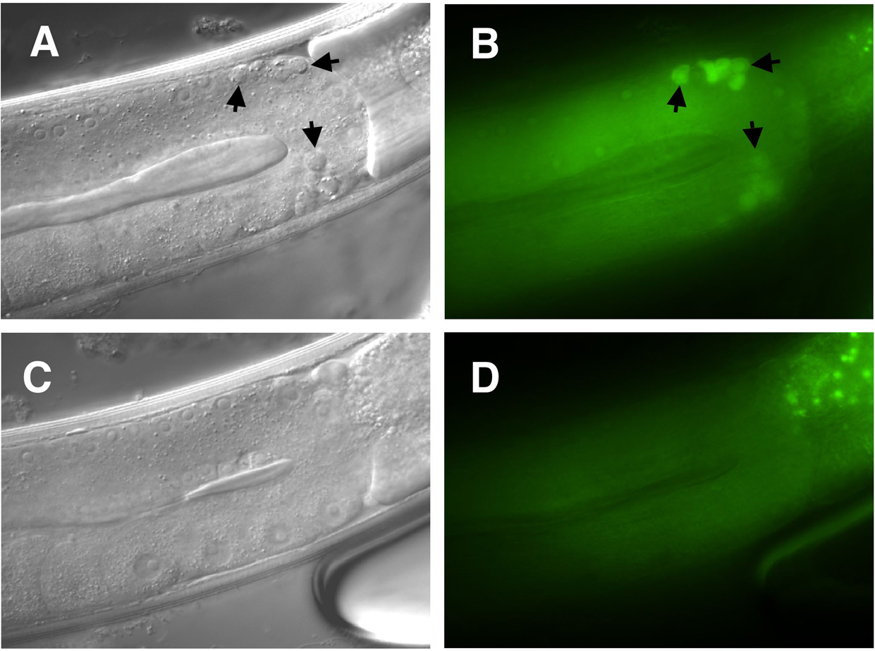
DAPI staining of mec-4(d) animals expressing pmec-17 LMP-1∷GFP. (A) ALM...  | Download Scientific Diagram
DAPI staining of mec-4(d) animals expressing pmec-17 LMP-1∷GFP. (A) ALM... | Download Scientific Diagram

Methods in cell biology

a) C. elegans adult hermaphrodite stained with DAPI to show nuclei;... |  Download Scientific Diagram
a) C. elegans adult hermaphrodite stained with DAPI to show nuclei;... | Download Scientific Diagram

Figures and data in Expansion microscopy of C. elegans | eLife

Figures and data in Expansion microscopy of C. elegans | eLife

Figure 4, Staining in wild-type versus transgenic strains - WormBook - NCBI  Bookshelf
Figure 4, Staining in wild-type versus transgenic strains - WormBook - NCBI Bookshelf

Methods in cell biology

DAPI staining of mec-4(d) animals expressing pmec-17 LMP-1∷GFP. (A) ALM...  | Download Scientific Diagram
DAPI staining of mec-4(d) animals expressing pmec-17 LMP-1∷GFP. (A) ALM... | Download Scientific Diagram

Superresolution microscopy reveals the three-dimensional organization of  meiotic chromosome axes in intact Caenorhabditis elegans tissue | PNAS
Superresolution microscopy reveals the three-dimensional organization of meiotic chromosome axes in intact Caenorhabditis elegans tissue | PNAS

Optical micrographs showing phase contrast images and DAPI fluorescence...  | Download Scientific Diagram
Optical micrographs showing phase contrast images and DAPI fluorescence... | Download Scientific Diagram

Expansion microscopy of C. elegans | eLife

Epifluorescence microscopic analysis of DAPI stained specimens. Many... |  Download Scientific Diagram
Epifluorescence microscopic analysis of DAPI stained specimens. Many... | Download Scientific Diagram

DAPI staining of wild-type Caenorhabditis elegans (Clade 9). The... |  Download Scientific Diagram
DAPI staining of wild-type Caenorhabditis elegans (Clade 9). The... | Download Scientific Diagram

Antibody Staining in C. Elegans Using "Freeze-Cracking" | Protocol

C. elegans embryo: Dapi, FiTC and Cy3 staining. widefield image,... |  Download Scientific Diagram
C. elegans embryo: Dapi, FiTC and Cy3 staining. widefield image,... | Download Scientific Diagram

Methods in cell biology

A,B) A wild-type C. elegans embryo was stained with DAPI (blue) and... |  Download Scientific Diagram
A,B) A wild-type C. elegans embryo was stained with DAPI (blue) and... | Download Scientific Diagram

DAPI staining of wild-type Caenorhabditis elegans (Clade 9). The... |  Download Scientific Diagram
DAPI staining of wild-type Caenorhabditis elegans (Clade 9). The... | Download Scientific Diagram

A,B) A wild-type C. elegans embryo was stained with DAPI (blue) and... |  Download Scientific Diagram
A,B) A wild-type C. elegans embryo was stained with DAPI (blue) and... | Download Scientific Diagram

Methods in cell biology

Light-microscopy methods in C. elegans research - ScienceDirect